This section is a review of basic shoulder anatomy. It covers the bones, ligaments, muscles and other structures that make up the shoulder. For more information on how the shoulder works please read the section on basic shoulder biomechanics.
A recommended book for learning shoulder anatomy is Moore’s Clinically Oriented Anatomy
Jmarchn, CC BY-SA 3.0 https://creativecommons.org/licenses/by-sa/3.0, via Wikimedia Commons
How many bones make up the shoulder complex?
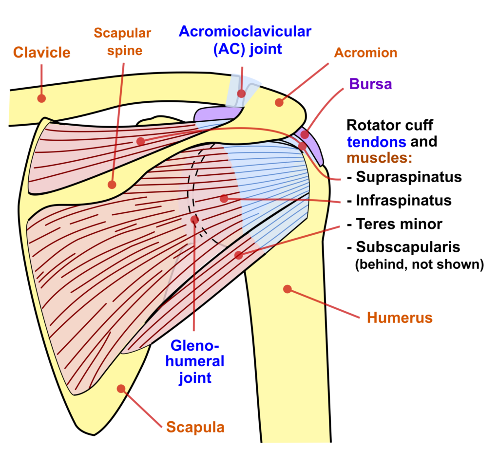
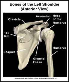
The shoulder complex is made up of three bones, which are connected by muscles, ligaments, and tendons. The large bone in the upper arm is called the humerus. The shoulder blade is called the scapula and the collarbone is called the clavicle. The top of the humerus is shaped like a ball. This ball sits in a socket on the end of the scapula. The ball is called the head of the humerus and the socket is called the glenoid fossa, hence the term “glenohumeral” joint. The glenoid fossa has a rim of tissue around it called the glenoid labrum. The glenoid labrum makes the glenoid fossa deeper. The glenohumeral joint is the most mobile joint in the body.
Articular cartilage is a smooth shiny material that covers the humeral head and the glenoid fossa of the glenohumeral joint. There is articular cartilage anywhere that the bony surfaces come into contact with each other. Articular cartilage allows these bones to slide easily over each other as the arm moves.
What are the joints that make up the shoulder complex?
The glenohumeral joint is just one of the joints in the shoulder complex. The other two joints are the sternoclavicular joint and the acromioclavicular joint. The sternoclavicular joint allows a small amount of movement to occur between the inner (medial) part of the clavicle and the breastbone (sternum). The acromioclavicular joint allows a small amount of movement to occur between the outer (lateral) part of the clavicle and a projection on the top of the scapula called the acromion process. The scapula sits on the back of the ribs and moves as the arm moves.
Ligaments are like strong ropes that help connect bones and provide stability to joints. In the shoulder complex, ligaments provide stability to the sternoclavicular joint, the acromioclavicular joint and the glenohumeral joint. The ligaments around the sternoclavicular joint and the acromioclavicular joint are strong and tight and do not allow for much movement in these joints.
The glenohumeral joint is surrounded by a large, loose “bag” called a capsule. The capsule has to be large and loose to allow for the many movements of this joint. Ligaments reinforce the capsule and connect the humeral head to the glenoid fossa of the scapula. These ligaments work with muscles to provide stability to the glenohumeral joint. The glenoid labrum also helps provide stability to the joint.
How many muscles surround the glenohumeral joint?
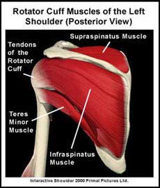
Tendons connect muscles to bone. There are four muscles (supraspinatus, infraspinatus, subscapularis and teres minor) that surround the glenohumeral joint. These four muscles are attached to the scapula. They turn into tendons, which in turn attach to the humerus. The tendons of these four muscles make up the “rotator cuff” that blends into and helps support the glenohumeral joint capsule. The muscles of the rotator cuff and their tendons provide stability to the glenohumeral joint when the arm is in motion. The biceps muscle is located in the front of the upper arm. It has two tendons, one of which attaches above the glenoid fossa. This tendon runs down the front of the glenohumeral joint and provides added stability to the glenohumeral joint.
There are muscles that stabilize the scapula and others that help move the arm. The rhomboid muscles, trapezius muscle and serratus anterior muscle are a few of the scapular stabilizing muscles. The pectoralis major muscle, the deltoid muscle and the muscles of the rotator cuff are some of the muscles that move the arm at the glenohumeral joint. The upper part of the trapezius muscle also helps “shrug” the shoulder. All of the muscles that are part of the shoulder complex work together in order to move the arm through its many possible ranges of movement.
Finally, a bursa (pl. bursae) is a fluid filled sac that decreases the friction between two tissues. Bursae also protect tissues from bony structures. In the shoulder, the subacromial bursa (also called the subdeltoid bursa) covers the rotator cuff tendons and protects them from the overlying acromion process. Normally, this bursa has very little fluid in it but if it becomes irritated it can fill with fluid, become painful and also irritate the surrounding rotator cuff tendons.
A recommended book for learning shoulder anatomy:
Moore’s Clinically Oriented Anatomy
Renowned for its comprehensive coverage and engaging, storytelling approach, the bestselling Moore’s Clinically Oriented Anatomy, 9th Edition, guides students from initial anatomy and foundational science courses through clinical training and practice. A popular resource for a variety of programs, this proven text serves as a complete reference, emphasizing anatomy that is important in physical diagnosis for primary care, interpretation of diagnostic imaging, and understanding the anatomical basis of emergency medicine and general surgery.