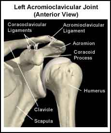
In order to understand osteolysis of the distal clavicle, sometimes called “Weightlifter’s Shoulder“, it is important to understand the anatomy and function of the shoulder. Please read Shoulder Pain Info’s section on basic shoulder anatomy and the anatomy of the acromioclavicular (AC) joint. For additional background information on the biomechanics of the shoulder please read Shoulder Pain Info’s section on basic shoulder biomechanics.
What is osteolysis?
The word “osteolysis” refers to a softening, absorption, and dissolution of bone or the removal or loss of calcium in bone. At the acromioclavicular joint the end of the clavicle can undergo osteolysis. Over time osteolysis of the end of the clavicle can result in the loss of 0.5 to 3 cm of bone.
What can cause AC joint osteolysis?
No one knows for sure what causes osteolysis of the distal clavicle but some risk factors include:
- A single injury to the AC joint or to the end of the clavicle
- Repetitive minor injuries to the AC joint or to the end of the clavicle
- Repetitive heavy weight lifting such as overhead shoulder press and bench press
- Pre-existing disease states such as rheumatoid arthritis, hyperparathyroidism, infection, multiple myeloma, and scleroderma.
What are the symptoms of osteolysis of the distal clavicle?
Osteolysis of the distal clavicle usually comes on slowly and results in shoulder pain, stiffness and/or swelling. The pain may is felt in the area of the AC joint or the end of the clavicle. The pain is usually made worse by activities such as bench press, shoulder press, push – ups and throwing.
Can osteolysis of the distal clavicle be detected on X-ray?
X-rays can be an effective tool for identifying osteolysis of the distal clavicle but the bony changes may take weeks or months before they can be seen on an X-ray. A bone scan is an effective tool to help identify early osteolysis. A bone scan will show increased uptake over the distal clavicle and, occasionally, increased uptake in the acromion process. Magnetic resonance imaging exhibits altered signal intensity in the distal clavicle but is not necessary to make a definitive diagnosis.
What is the treatment for osteolysis of the distal clavicle?
The goal of treatment of osteolysis of the distal clavicle is to reduce pain while the clavicle “remineralizes”. Rest or activity modification, anti-inflammatory medications and ice are usually prescribed to reduce pain. If these measures are not effective an injection of cortisone into the AC joint may be necessary. In most cases, the clavicle slowly remineralizes (over 4 to 6 months), but may take on a tapered appearance. In some cases, the bones do not remineralize and a surgical consult may be required. The surgeon may consider resecting (removing) part of the affected clavicle to reduce symptoms.
Can osteolysis of the distal clavicle be prevented?
When symptoms of AC joint pain first develop, avoiding pain provoking activities is recommended. Additional padding for contact sports can also be effective. Finally, weight lifters should avoid locking their elbows during the bench press, use a narrower grip on the bar, and avoid bending their elbows past horizontal.
What other information is available on osteolysis of the distal clavicle?
Shoulder Pain Info’s links section has additional information on this topic. Links have been provided to other websites as well as online medical journals.