In order to understand this condition, it is important to understand the anatomy and function of the shoulder. Please read Shoulder Pain Info’s section on basic shoulder anatomy. For additional background information on the biomechanics of the shoulder please read Shoulder Pain Info’s section on basic shoulder biomechanics.
What is important to know about the anatomy of the shoulder complex?
The shoulder complex is made up of three bones, which are connected by muscles, ligaments, and tendons. The large bone in the upper arm is called the humerus. The shoulder blade is called the scapula and the collarbone is called the clavicle. The top of the humerus is shaped like a ball. This ball sits in a socket on the end of the scapula. The ball is called the “head” of the humerus and the socket is called the glenoid fossa, hence the term “glenohumeral” joint. The glenoid fossa has a rim of tissue around it called the glenoid labrum. The glenoid labrum makes the glenoid fossa deeper. The glenohumeral joint is the most mobile joint in the body.
The glenohumeral joint is surrounded by a large, loose “bag” called a capsule. The capsule has to be large and loose to allow for the many movements of this joint. Ligaments reinforce the capsule and connect the humeral head to the glenoid fossa of the scapula. These ligaments work with muscles to provide stability to the glenohumeral joint. The glenoid labrum also helps provide stability to the joint.
Mikael Häggström, M.D., CC0, via Wikimedia Commons
What is a shoulder dislocation?
A shoulder dislocation occurs when the head of the humerus moves out of it’s normal position in the socket of the shoulder complex (the glenoid fossa). As a result the ligaments that stabilize the glenohumeral joint are injured or torn.
There are different types of shoulder dislocations. Classification of the different types depends on the direction in which the head of the humerus dislocates. The most common type of shoulder dislocation is one where the head of the humerus dislocates in front of and below the glenoid fossa. This is called and anterior (in front of) – inferior (below) shoulder dislocation.
Anterior-inferior shoulder dislocations make up over 95% of shoulder dislocations. For this reason, the rest of this article will only be discussing anterior-inferior shoulder dislocations.
What causes an anterior-inferior shoulder dislocation?
Anterior-inferior shoulder dislocations usually occur when there is contact with the arm while the arm is away from the body in an outward rotated position. Traumatic contact from the back of the shoulder can also cause this type of dislocation even when the arm is by the side of the body. These traumatic events will cause the head of the humerus to be displaced out of the glenoid fossa.
What does a shoulder dislocation feel like?
Immediately following a shoulder dislocation there is usually pain in the area around the shoulder. While the shoulder is dislocated, trying to move the arm or shoulder will usually increase the pain. There is usually some visible displacement of the head of the humerus anterior and inferior to the glenoid fossa and as a result the injured shoulder does not look the same as the other shoulder. Sometimes certain parts or all of the shoulder or the arm may feel numb.
What are some possible complications of an anterior-inferior shoulder dislocation?
- Increased tendency to have more shoulder dislocations in the injured shoulder.
- Tears of the rotator cuff.
- Fractures (broken bones) of the head of the humerus or of the glenoid fossa.
- Fractures of other areas of the upper arm bone (the humerus).
- Reversible or irreversible nerve damage.
- Reversible or irreversible blood vessel damage.
- Frozen shoulder.
Mikael Häggström, M.D. – CC0, via Wikimedia Commons
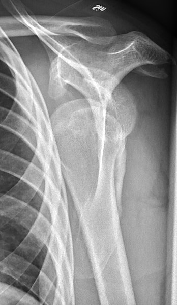
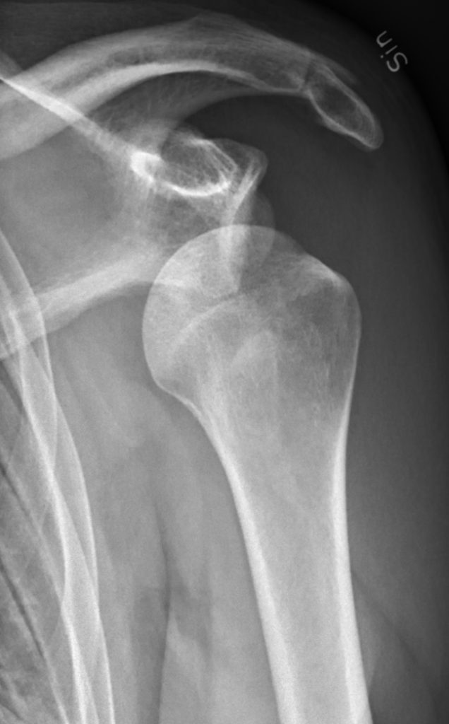
Can an anterior-inferior shoulder dislocation be detected on X-rays?
X-rays are an effective tool for identifying anterior-inferior shoulder dislocations and they are also used to identify when the head of the humerus has returned to its proper position. X-rays are also very effective for identifying fractures.
X-rays are usually taken when a shoulder dislocation is first suspected and after the head of the humerus has been returned to it’s proper position to make sure that it is in the right place.
Certain special tests can be performed by a physician or physical therapist to diagnose instability in the shoulder that could indicate a previous shoulder dislocation. Such special tests of the shoulder include:
- Apprehension Test
- Crank Test
- Hyper Extension-Internal Rotation (HERI) Test
- Jobe Relocation Test
- Sulcus Sign
What is the treatment for an anterior-inferior shoulder dislocation?
Usually, anterior-inferior shoulder dislocations are treated by doctors, often in the emergency department. Doctors trained in treating emergencies are also trained in making sure that the head of the humerus is put back into it’s correct position. Sometimes the head of the humerus does not want to return to it’s correct position and an Orthopedic Surgeon is consulted on whether surgery is required.
After an anterior-inferior shoulder dislocation the long term goal is to return the individual back to their previous level of activity. Achieving this goal will depend on the function and stability of the shoulder and any complications that have occurred. After a period of immobility (note that sometimes immobilizing the shoulder is not recommended), a general shoulder rehabilitation program which includes strengthening exercises, flexibility exercises, aerobic conditioning, technique refinement and proprioceptive (biofeedback) retraining is the most important factor in improving shoulder function and stability. Stability may be improved by an anterior-inferior shoulder dislocation brace. If the shoulder remains unstable or if an individual has repeated anterior-inferior shoulder dislocations, surgery to stabilize the shoulder is often required.
What other information is available on anterior-inferior shoulder dislocations?
Shoulder Pain Info’s links section has additional information on this topic. Links have been provided to other websites as well as online medical journals.